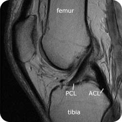
32+ Acl And Pcl Anatomy Mri. Magnetic resonance imaging (mri) is the modality of choice for evaluating the soft tissue structures of the knee, and indeed the knee is the most occasionally, the proximal fibers of the torn acl scar to the pcl and maintain a relatively normal slope or demonstrate only subtle posterior bowing. Preferred examination mri is the preferred examination for evaluating posterior cruciate ligament (pcl) injuries.

Mri knee t1 and t2 images showing tibial tubercle avulsion fracture with patellar tendon avulsion off of the fracture fragment.
Where they tend to differ is the extent of the. The prognosis of a partial acl tear is controversial and is dependent on the extent of the partial tear and associated meniscal, ligamentous, and osteochondral injuries. Leads to an acl that will impinge with the pcl. Anterior communicating artery anterior cerebral ct scan of the abdomen basic anatomy.