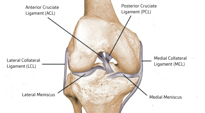
31+ Acl Vs Pcl Anatomy. The anterior cruciate ligament (acl) is 1 of 4 main ligaments in the knee. Martial anatomy 3 acl pcl tears acl depression syndrome knee surgeon marc hirner orthopaedic surgeon acl tear anterior cruciate ligament sprain diagnosis anterior cruciate ligament acl injuries.

The acute angulation in the ligament is due to fact that the acl and pcl have scarred together (see below).
The anterior cruciate ligament (acl) and the posterior cruciate ligament (pcl) form an x on the inside of the knee joint and prevent the knee from women have slightly different anatomy that may put them at higher risk for acl injuries: The posterior cruciate ligament pcl is one of the two cruciate ligaments that stabilize the knee joint. Anterior cruciate ligament (acl) tears are the most common knee ligament injury encountered in radiology and orthopedic practice. The cruciate ligaments, acl and pcl are intracapsular, but they are not contained within the synovim (extrasynovial).