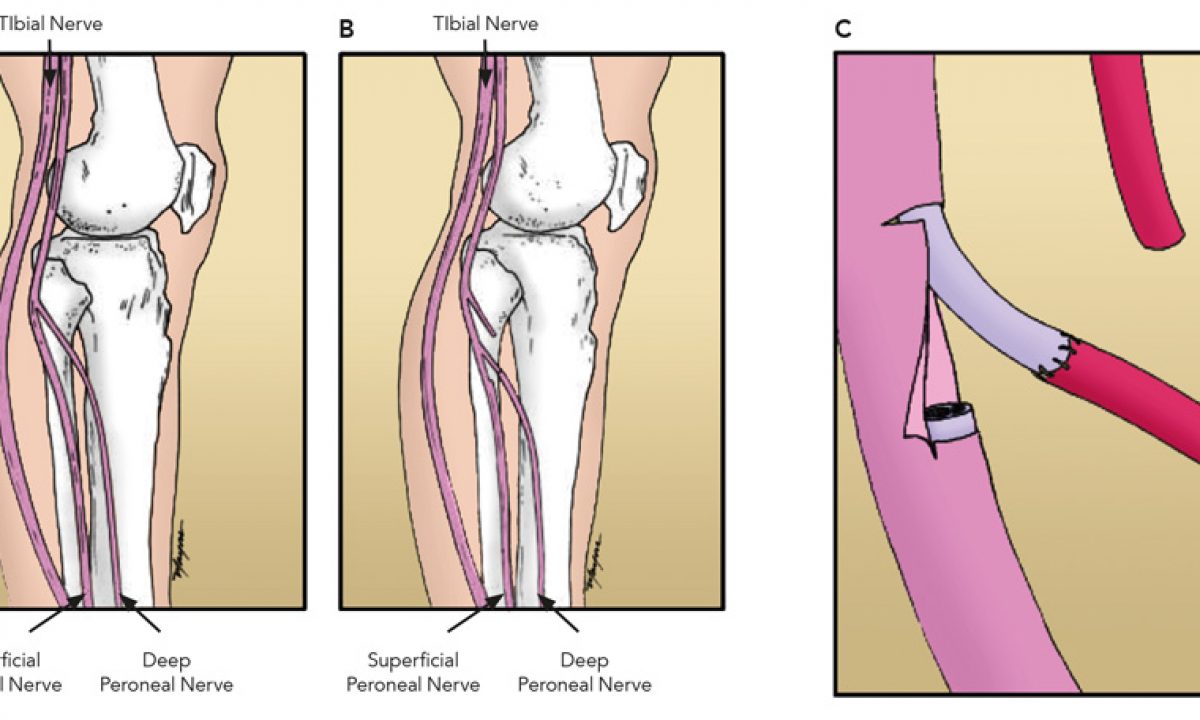
50+ Peroneal Nerve Anatomy Knee. The common peroneal nerve, also known as common fibular nerve, forms the lateral part of the sciatic nerve and supplies the leg. The common peroneal nerve starts just above the knee and supplies sensory and motor function to the lower leg and foot.

Here we discuss the anatomy of the deep peroneal nerve (aka deep fibular nerve) including deep peroneal nerve:
The nerve then dives into the peroneus longus muscle, where tethering can occur, making it susceptible to stretch injury at this level. The common peroneal nerve, which is also known as the common fibular nerve, provides sensory innervation to the inferior portion of the knee joint, and the posterior and lateral skin of the upper calf. The common peroneal nerve travels laterally and courses around the fibular neck and passes through an opening in the peroneus. Normal anatomy and pathologic findings on routine mri of the knee.