Muhammad bin zulfiqar pgr iii fcps new radiology department services hospital / services institute of medical 15.
41+ Sagittal Mri Shoulder Anatomy. Normal anatomy, variants and checklist. Notice rotator cuff muscles and look for atrophy.
Mri Of The Shoulder from image.slidesharecdn.com
In the coronal oblique plane, slices are made parallel to the tendon of the supraspinatus muscle, starting from the posterior to the. For an accurate evaluation is essential to know the normal anatomy and variants to avoid the misdiagnosis of a pitfall as pathological condition. In this article, we shall look at the anatomy of the shoulder joint and its important clinical correlations.
Normal anatomy, variants and checklist.
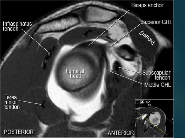
Robin smithuis and henk jan van der woude. This mri shoulder cross sectional anatomy tool is absolutely free to use. Highest section shows supraspinatus muscle followed posteriorly by infraspinatus muscle & teres minor & its tendons. The use of mr for the diagnosis of shoulder lesions is well established, as well as mr arthrography for the diagnosis for shoulder instability and microinstability.