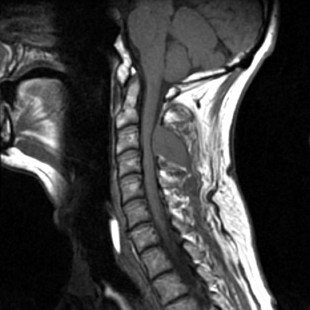
46+ C Spine Anatomy Mri. The video describes the cervical spine anatomy and the approach to reading a cervical spine mri by dr. This webpage presents the anatomical structures found on knee mri.

Nasopharynx, orbit, paranasal sinuses, cranial nerves, temporomandibular joint, neck, brachial plexus, spine, shoulder, arm, elbow, forearm, wrist.
Further imaging with ct or mri (not discussed) is often appropriate in the context of a high risk injury, neurological deficit, limited clinical examination tap on/off image to show/hide findings. Athletic injuries to the head, neck, and face. Anatomy for diagnostic imaging 2e. Imaging should provide enough anatomic coverage to detect transitional anatomy at the lumbosacral junction.