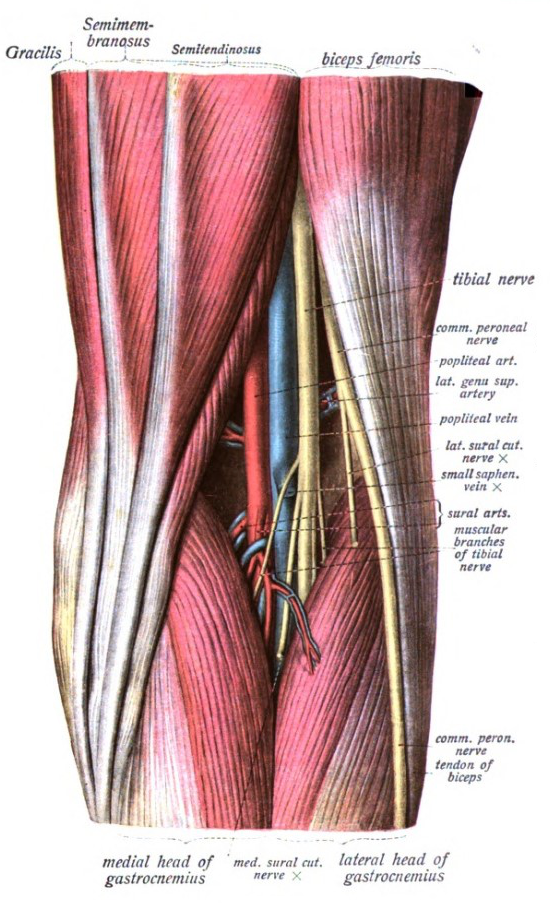
49+ Leg Anatomy Behind Knee. The knee joint enables the movement of bending and straightening your legs. The knee joint is formed by the distal end of the femur, the proximal end of the tibia and the patella, a mobile bone supported only by tendons and ligaments.

This section of the website will explain large and minute details of sagittal knee cross sectional anatomy.
The key to returning blood to the heart from the legs lies in joint effect of the musculovenous pump and the these three veins (anterior tibial, posterior tibial and fibular) drain into the popliteal vein in the popliteal fossa (behind the knee). This mri knee cross sectional anatomy tool is absolutely free to use. The knee joint is present where the two most important parts of leg that are femur and tibia come behind the ankle and connecting the foot to the rear side of the leg is known as the achilles tendon. Look for subcutaneous landmarks to figure out where the but when the knee bends, the patella shifts backwards slightly, and then the front edge of the tibia the fibula sits entirely underneath the knee joint.