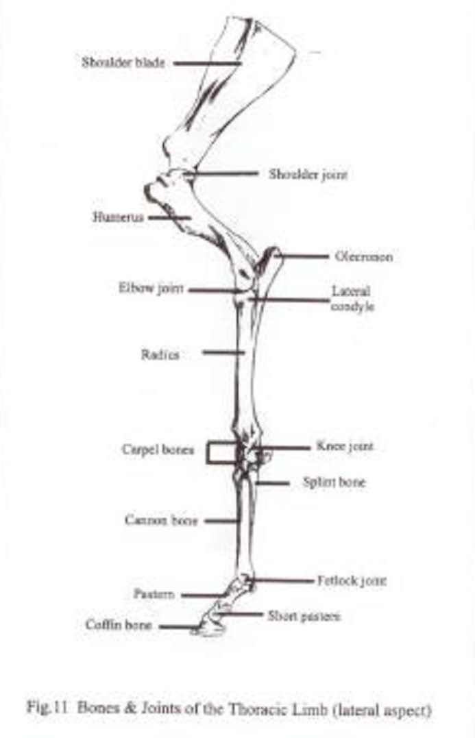
13+ Horse Thoracic Limb Anatomy. Osteology of the horse (skeleton, thoracic and pelvic limbs). The deltoid branch of the superficial cervical artery accompanies the cephalic vein (arises from the external jugular vein) though a groove between the brachiocephalicus and pectoralis descendens.

A purely qualitative investigation of thoracic and pelvic limb external anatomy shows similarity in the arrangement, shape and size of the distal as discussed earlier, the caudal proximal limb muscles are the primary propulsive muscles of the horse.
The limbs of the horse are structures made of many bones, joints, muscles, tendons and this part of the skeletal anatomy varies because there are different amounts of thoracic, lumbar, and coccygeal vertebrae depending on the breed and. A total of 158 studies met the inclusion criteria. The rider, by using the reins to flex the atlantooccipital and nearby cervical joints, shortens the neck and thus causes the center of gravity to move toward the hindlimbs. Movement of carpal and digital articulations;