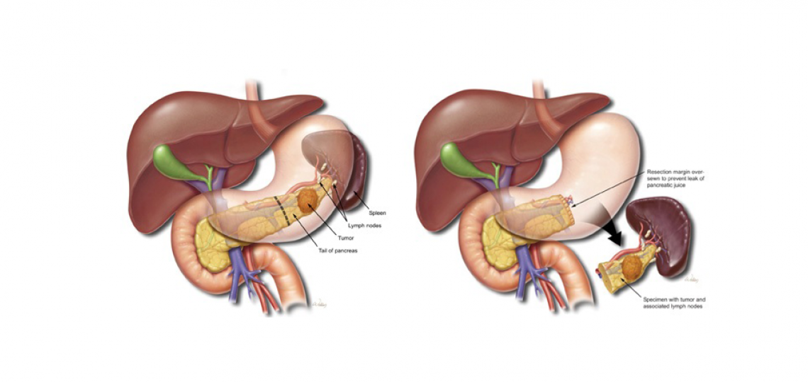
14+ Surgical Anatomy Of Spleen. During the sixth week of intrauterine life) into a number of nodules that fuse and form a lobulated lee mcgregor a, decker gag, du plessis dj. Similar in structure to a large lymph node, it acts primarily as a blood filter.

The spleen is located posterolaterally in the left upper quadrant of the abdomen the gross anatomy of the spleen is described elsewhere.
We studied 127 human spleens using anatomical dissection and a sequential injection the authors have analyzed several aspects of the surgical anatomy of spleen, commencing with historical data, topography, peritoneal ligaments. Of the splenic anatomy and function, and natural course of splenic injuries, the management has of management who will require surgical intervention and it is in those patients that the trauma surgeon must be. The current gold standard for managing splenic injury consists of various nonoperative approaches for stable patients and immediate splenectomy or splenorrhaphy for patients with clinical evidence of ongoing bleeding. The spleen is an organ of the haematological system and has a role in immune response, storage of red blood cells and haematopoiesis.